Figure 1.
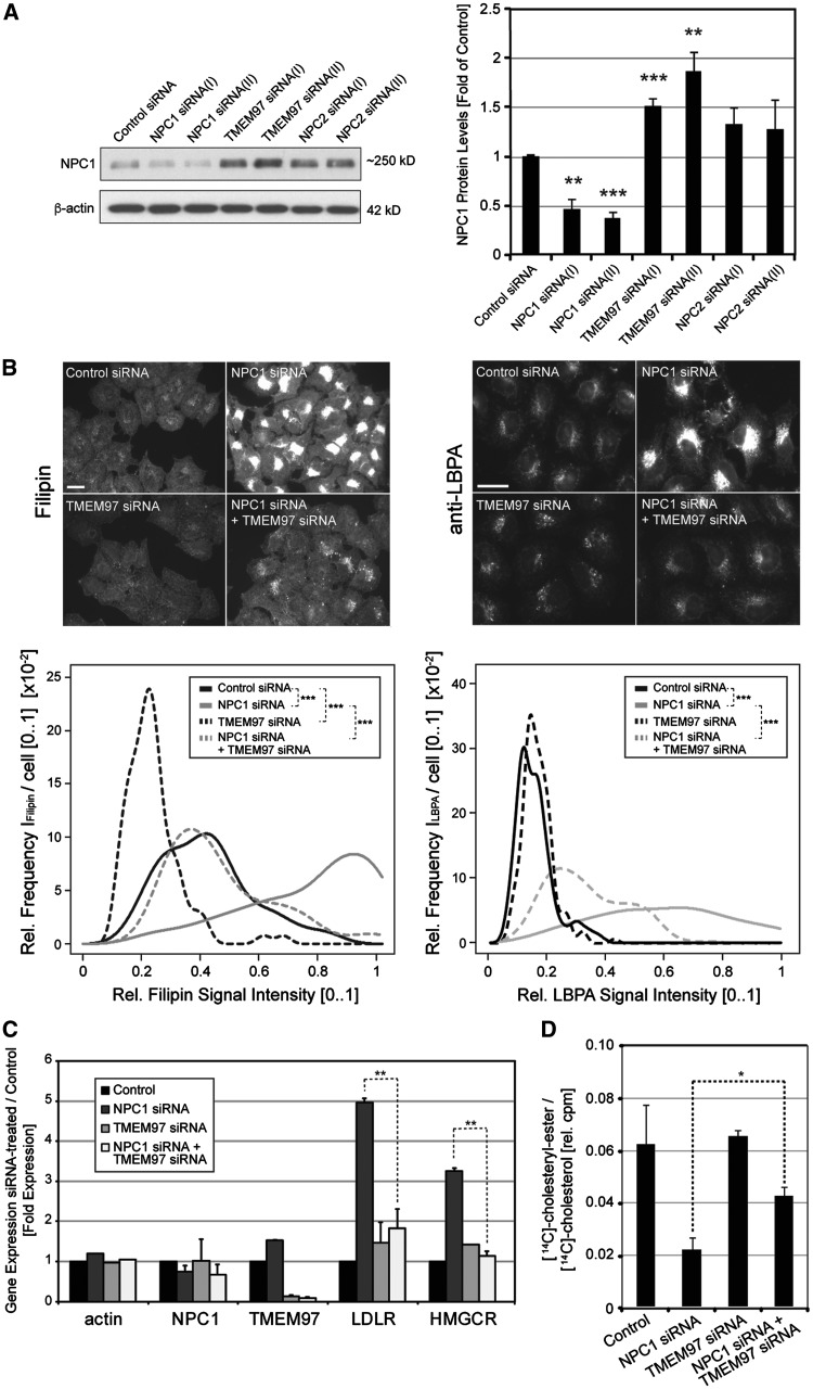
SiRNA-mediated knockdown of TMEM97 increases NPC1 protein levels, ameliorates cholesterol accumulation and restores cholesterol delivery to the ER in NPC1-deficient HeLa cells. (A) Cultured HeLa cells were transfected for 48h with either control siRNA or indicated siRNAs targeting NPC1, TMEM97 or NPC2. Whole cell lysates were subjected to Western blotting and probed with antibodies against NPC1 and β-actin. NPC1 protein levels were quantified as a ratio to β-actin and normalized to levels of control siRNA treated cells (n = 3 independent experiments per condition; **P < 0.01; ***P < 0.001). (B) HeLa cells transfected with siRNAs targeting indicated genes for 48h were stained with either filipin dye (left panel) or a polyclonal antibody against LBPA (right panel) (scale bars = 10µm). Filipin and LBPA signal intensities in perinuclear areas were quantified as the relative intensity frequency distributions [0.1] (y-axis) of mean perinuclear filipin or LBPA signals per cell (x-axis) from up to 20 images per condition with an average of 25 cells per image (n = 2–3 independent experiments per condition, ***P < 0.001). (C) HeLa cells cultured in DMEM/5%FCS were transfected with siRNAs against the indicated genes. Eight hours after siRNA-transfection, medium was changed to DMEM/0.5%LDS for 16h. Next, cells were washed and medium was changed to DMEM/10%FCS/50μg/ml LDL for another 24h before mRNA was extracted. Mean mRNA-levels of indicated genes relative to RPL19 are shown (n = 2–3 independent experiments per condition; **P < 0.01). (D) Analysis of cholesteryl-ester formation from LDL-associated [14C]-cholesterol in HeLa cells transfected with indicated siRNAs. 24h after siRNA-transfection, medium was changed to DMEM/0.5%LDS and cells were cultured for another 24h. Next, pulse-labeled medium containing DMEM/0.5%LDS/50μg/ml LDL was added and [14C]LDL-cholesterol was internalized for 4.5h before cellular lipids were extracted and separated by thin-layer chromatography. Cell-associated free [14C]-cholesterol and [14C]-cholesteryl-ester signals (in counts per minute, cpm) were quantified by scintillation counting (n = 2 independent experiments; *P < 0.05).

