Figure 2.
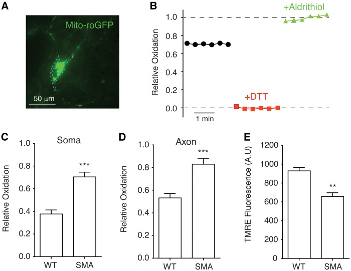
Increased mitochondrial oxidative stress and compromised membrane potential in spinal motor neurons affected by SMA. (A) A primary mouse spinal motor neuron expressing redox-sensitive green fluorescent protein with mitochondria targeting sequence (mito-roGFP). The contour of motor neuron was marked by dashed lines. (B) Measurement of mitochondrial oxidation level. The intensities of mito-roGFP in motor neurons were measured with a live imaging fluorescent microscope. DTT were applied to fully reduce (red trace) and aldrithiol were used to fully oxidize (green trace) roGFP for calculating relative oxidation levels as described in Methods. (C, D) Mitochondrial oxidative stress was significantly increased in both the soma (C) (P = 0.00013) and axon (D) (P = 0.0003) of spinal motor neurons from Δ7 SMA mice. Mitochondrial roGFP oxidation levels in the soma and axon (at least 50 µm away from the motor neuron soma) were recorded in 5–7 experimental repeats for each group. (E) Mitochondrial membrane potential was significantly reduced (P = 0.0069) in SMA motor neurons. The membrane potentials of mitochondria on 42 samples of Δ7 SMA motor neurons and 42 wild type samples in three independent experiments were measured using fluorescent dye tetramethylrhodamine ethyl (TMRE) and normalized to cell number. **P < 0.01; ***P < 0.001, Student’s t test.

