Figure 4.
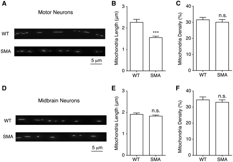
Mitochondria fragmentation in spinal motor neurons from Δ7 SMA mice. (A) A representative image for measuring mitochondria length and density in wild type (WT) and SMA mouse motor neuron axons. Primary spinal motor neurons were transfected with mito-dsRed at 5-7 days in vitro and imaged using a confocal microscope 48 h later. Mitochondria length and density were analysed along axons at least 50 µm away from the soma. (B, C) Mitochondria length in spinal motor neurons from SMA mice was significantly shorter (P = 0.00001), than that of wild type motor neurons while mitochondria density was not changed (P = 0.51). Wild type motor neurons: 174 mitochondria from 40 axons; SMA motor neurons: 260 mitochondria from 58 axons. (D) A representative image of mitochondria in wild type and SMA mouse midbrain neuron axons. (E, F) Neither mitochondria length (E) (P = 0.33) nor density (F) (P = 0.53) was changed in SMA midbrain neurons. Wild type midbrain neurons: 316 mitochondria from 23 axons; SMA midbrain neurons: 378 mitochondria from 33 axons. Results are mean ± SEM, from at least three different cultures. n.s: not significant; ***P < 0.001, Student’s t test.

