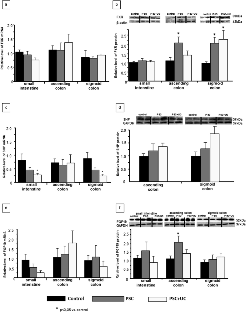
Figure 2. Intestinal expressions of FXR, SHP, and FGF19.
Adaptive regulation of FXR expression was evaluated in three parts of the gut, with comparison between cases of cholestasis and control tissues. (a) FXR mRNA expression was comparable between the terminal ileum, ascending colon, and sigmoid colon in all groups. (b) FXR protein expression did not differ between PSC patients and controls in the ileum, but was significantly enhanced in PSC patients compared to control subjects in both the ascending and sigmoid colon. Within the PSC + UC group, this increased FXR protein expression was only detected in the sigmoid colon. (c) SHP mRNA expressions in the terminal ileum and the colon were equal among all examined groups, aside from a lower SHP mRNA level in the descending colon among PSC + UC subjects. (d) SHP protein levels were similar among all analysed samples regardless of the part of the gut. (e) FGF19 mRNA levels were similar in all examined parts of the gut, regardless of the examined group. (f) Intestinal FGF19 protein levels in PSC and PSC + UC patients were comparable to in controls, except for an elevated level in the ascending colon of PSC patients. mRNA expression levels are presented as fold-change relative to control, and were normalized relative to glyceraldehyde 3-phosphate dehydrogenase (GAPDH). Changes in protein levels were determined using densitometry analyses following normalization to GAPDH or β-actin as a loading control, and are presented as fold-change relative to control.

