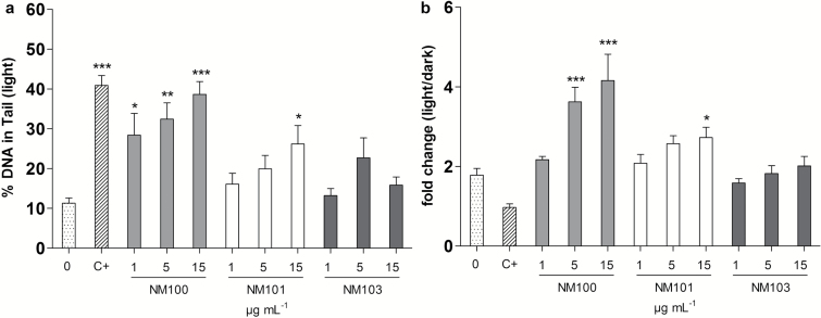
Figure 6.
Additional DNA strand breaks caused by light. BEAS-2B cells were exposed to particles for 24h, and comet slides were exposed to normal lab light for 3min + 3min (after lysis and alkaline treatment, respectively). Results are presented as mean ± SEM (a) and fold change compared to dark conditions (as shown in Figure 5) (b). *P < 0.05, **P < 0.01, ***P < 0.001. C+, 2 µM Ro together with light irradiation.

