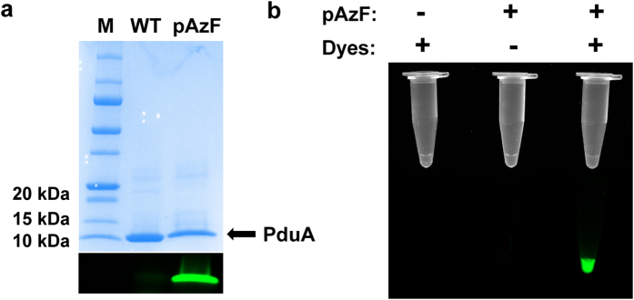
Figure 2. Incorporation of p-azido-phenylalanine (pAzF) in Salmonella PduA protein.
(a) The SDS-PAGE gel of PduA labeling. The upper panel is the gel stained with Coomassie Blue. The lower panel was captured with fluorescent filters for Alexa Fluor 488. (b) The fluorescence labeling of purified Pdu microcompartments in vitro. The upper panel was captured without filters. The lower panel was captured with fluorescent filters for Alexa Fluor 488. The left lane was wild-type Pdu MCPs with dyes. The middle lane only contained pAzF-containing Pdu MCPs. The right lane included both pAzF-containing Pdu MCPs and dyes.

