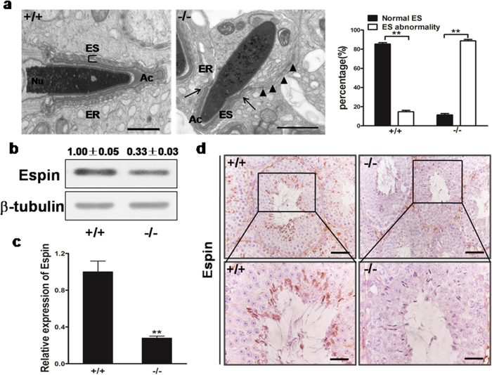
Figure 6. Analysis of Mkrn2 knockout mouse ectoplasmic specialization (ES) structure.
(a) TEM analysis of ES structure. Arrows indicate the boundary of the acrosome, and arrowheads indicate the improper arrangement of ES. Nu, nucleus; Ac, acrosome; ER, endoplasmic reticulum. Data are represented as means ± SE. From six different testis tissues for each phenotype. Error bars represent SE. **P < 0.01 (two-tailed Student’s t test). Scale bar = 1 μm. (b) Espin expression levels in the testes were analyzed by immunoblotting. β-Tubulin was used as the internal control. (c) The Espin mRNA levels in the testes were analyzed by RT-qPCR and normalized to that of β-Tubulin. Data are represented as means ± SE. From six different testis tissues for each phenotype. Error bars represent SE. **P < 0.01 (two-tailed Student’s t test). (d) Immunohistochemical staining of Espin in the testes shows co-localization of Espin at the apical ES and in the heads of elongated spermatids in wild-type testes (left), but no obvious staining can be observed in the tubules of the Mkrn2 knockout testes. Scale bar: Upper panel, 50 μm; Bottom panel, 20 μm.

