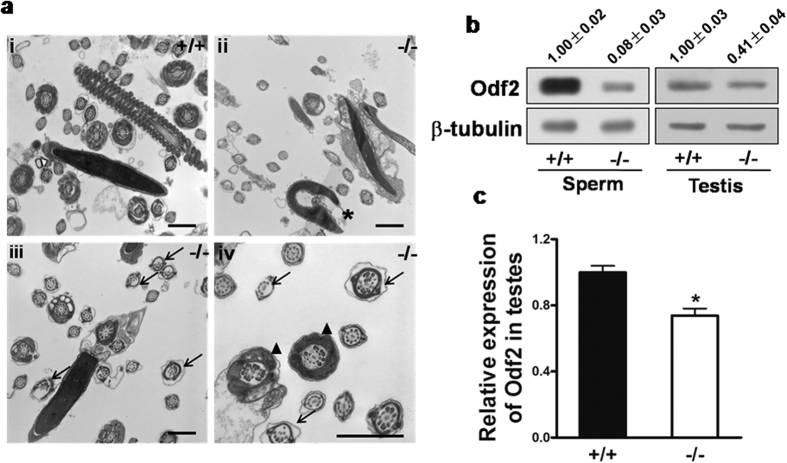
Figure 7. Analysis of Mkrn2 knockout mouse sperm structures and Odf2 expression.
(a) TEM analysis of epididymal sperm. i, Note the normal head and axoneme with typical “9 + 2” microtubule structure (nine pairs of peripheral and two central microtubules) in wild-type mice. ii–iv, Abnormalities in Mkrn2 knockout mouse sperm were evaluated by the appearance of deformed heads (ii, asterisk), deformed “9 + 2” structures with missing and/or misarranged microtubules (iii-iv, arrows) and lack of Odf (iv, arrowhead). Scale bar = 1 μm. (b) The Odf2 protein levels in wild-type and Mkrn2 knockout mouse sperm and testes, respectively. β-Tubulin was used as the loading control. (c) The Odf2 mRNA expression levels in the testes were determined by RT-qPCR and normalized to that of β-Tubulin. Data are represented as the means ± SE. From six different testes tissue samples for each phenotype. Error bars represent SE. *P < 0.05 (two-tailed Student’s t test).

