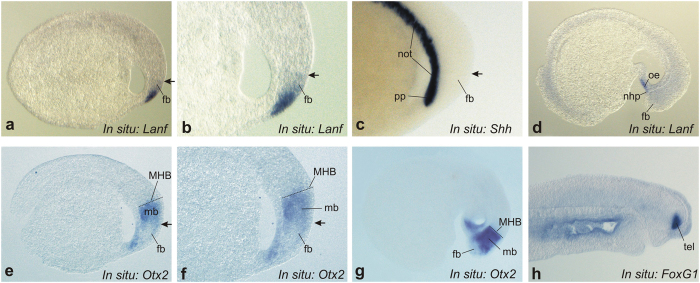
Figure 3. Analysis of the spatial expression pattern of Lanf in sagittal sections of L. fluviatilis embryos.
(a–c) At stage 19, Lanf ((a) - overall view, (b) - enlarged view of anterior region) is expressed in the anterior neurectoderm, in the region corresponding to the stomodeal ectoderm (se) and the forebrain (fb), including the presumptive telencephalon and diencephalon. This region is located just above the region of Shh expression in the notochord (not) and the prechordal plate (pp) (c). An arrow indicates the dorsal limit of Lanf expression. Anterior is to the right; dorsal is at the top. (d) At stage 23, Lanf expression is observed only in the stomodeal ectoderm (se) and the nasohypophysial placode (nhp). (е,f) At the same stage shown for Lanf in (a,b), Otx2 is expressed in a much broader area of the anterior neurectoderm, up to the presumptive mid-hindbrain boundary (MHB), and in the underlying mesoderm. Notably, the most anterior region of the neurectoderm, in which Lanf is expressed, is free of Otx2 expression. (g) At stage 23, Otx2 continues to be expressed in the anterior region, being inhibited in the most rostral part of it. (h) The expression of FoxG1 marks the telencephalon beginning from stage 25 (see Materials and Methods).

