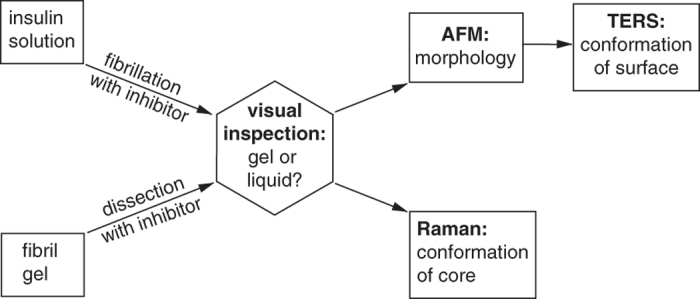
Figure 2. Analytical procedure for the characterization of all samples obtained from fibrillation and dissection experiments.

The first step is a simple visual inspection of the sample, followed by AFM and TERS experiments to characterize the morphology and structural conformation of the surface of single particles. Finally, conventional Raman measurements of the bulk sample allow the characterization of the fibril’s core secondary structure.
