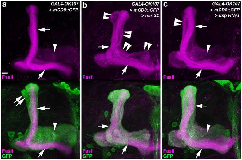
Figure 1. Ectopic miR-34 overexpression caused aberrant lobe formation on mushroom body (MB) neurons.
Confocal images show the lobe phenotype of mushroom body (MB) neurons in wild-type flies (a) and flies with ectopic miR-34 overexpression (b) and RNAi knockdown of USP (c). Fasciculin II (FasII) staining (magenta) reveals the dorsal α and medial β lobes (arrows, strong magenta staining) and the medial γ lobes (arrowheads, faint magenta staining) on MB neurons. In the lower panels, GAL4-OK107-driven mCD8::GFP (GFP) expression (green) shows the morphology of α lobes (arrows), α′ lobes (double arrows), and γ lobes (arrowheads). (a–c) Compared to the α and β lobes of wild-type MB neurons (arrows, a), ectopic miR-34 expression resulted in aberrant axonal branches projecting adjacent to the α and β lobes (double arrowheads, b). This miR-34 induced lobe defect is similar to that of the γ lobe phenotype observed in MB neurons in which USP expression was knocked down by RNAi (double arrowhead, c). Fly genotypes are listed in Supplemental Table 2. Scale bar: 10 μm for all panels.

