
Fig. 7.
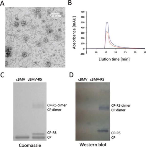
Biochemical characterization of cBMV–R5. (A) Transmission electron micrograph of cBMV–R5. (B) Size exclusion chromatogram of cBMV–R5 monitored at 260 nm (blue), 280 nm (red) (Abs 260 : 280 nm = 1.7). Intact cBMV–R5 fractions were collected and further characterized. (C) SDS-PAGE of cBMV and cBMV–R5 visualized under white light after Coomassie staining. Modified coat proteins (CP–R5) have an increased molecular weight. The ratio between CP–R5 : CP was determined based on band intensity and found to be 1 : 3. (D) Western blot probed with streptavidin–alkaline phosphatase detecting the biotin-tag of the R5 peptide.