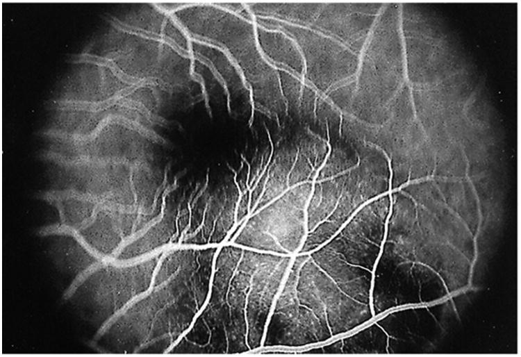
Figure 12.

Fluorescein angiogram in the early venous phase showing early blockage at the edges of the lesion and early hyperfluorescence within the central aspect of the choroidal lesion; the overlying retinal vessels are normal and in focus. The other retinal vessels are not in focus secondary to the thickness of the lesion. Reprinted with permission from the Archives of Ophthalmology (117). Copyright 2000 American Medical Association. All rights reserved.
