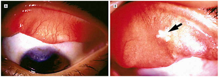
Figure 3.

(A) Case 1. At 1-year follow-up, the left eye shows a superior vascularized corneal scar with normal-appearing bulbar and tarsal conjunctiva. (B) Case 2. At 3-month follow-up, the everted right upper eyelid shows a residual area of necrosis (arrow) with mild persistent papillary reaction. Reprinted with permission from the Archives of Ophthalmology (27). Copyright 2003 American Medical Association. All rights reserved.
