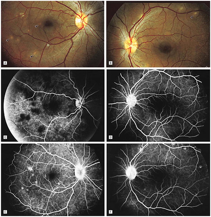
Figure 8.

Fundus photographs of the right (A) and left (B) eyes show bilateral, multifocal choroiditis (arrowheads). Serial FA photographs (C to F) show early blocking hypofluorescence and late-staining hyperfluorescence corresponding to areas of choroidal infiltrate, as well as mild, late leakage from the optic nerve heads in each eye. Reprinted with permission from the Archives of Ophthalmology (53). Copyright 1998 American Medical Association. All rights reserved.
