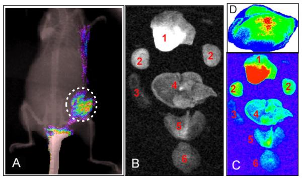
Figure 3.

(A) The whole-body NIR fluorescence image overlaid on an x-ray image showed the presence of (GdDOTA)42-G4-DL680 in the flank tumor of MDA-MB-231, as indicated dotted circle. (B and C) Ex-vivo light and fluorescence imaging of various organs of a mouse. Distribution of nanoparticles in organs: Light (B) and NIR (C) fluorescence images of tumor (1), kidney (2), spleen (3), liver (4), heart and lung (5) and brain (6) obtained from a mouse bearing a tumor after 24 post injection of (GdDOTA)42-G4-DL680 agent. (D) Ex-vivo fluorescence imaging of the flank tumor section (15 micron thick frozen flank tumor tissue section) clearly showed the accumulation of (GdDOTA)42-G4-DL680 in the tumor selectively. Non-specific accumulation of the particles was not observed.
