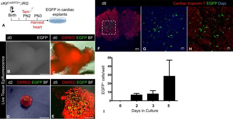
Figure 1. Neonatal heart cells originally marked by cKitCreERT2/+ are non-contractile and proliferate.
A, Schematic of the ex-vivo genetic fate-mapping strategy to assess the original identity of cKitCreERT2/+ -recombined heart cells. B-C, Live-tissue fluorescence imaging of tamoxifen-pulsed neonatal cardiac explants. At the time of harvest [Day (d)0], explants are DSRED+ and do not express EGFP epifluorescence. D-E, Live-tissue fluorescence imaging of tamoxifen-pulsed neonatal cardiac explants on d2 (D) and d5 (E) of culture. Expression of EGFP is restricted in a minor population of non-contractile cardiac cells, which proliferate with time. F-H, Confocal immunofluorescence against cardiac troponin T and EGFP of PN1 explants, after 8 days in culture. Panels G-H are a higher magnification of the area in inset of panel F. EGFP does not co-localize with cyanine-5 labeled cardiac troponin T. I, Quantification of EGFP+ cells during a 5-day culture period. BF, Brightfield. Scale bars, B-E, 200μm; F, 100μm; G-H, 20μM.

