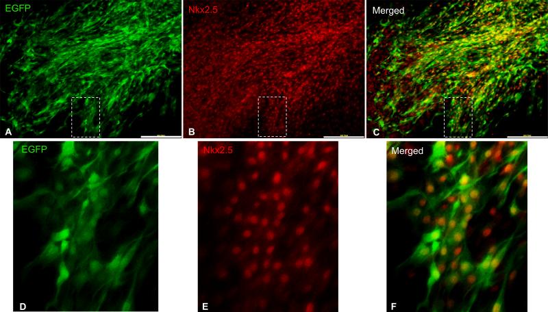
Figure 3. Co-localization of EGFP and Nkx2.5 in cardiac explant derived cells from cKitCreERT2;IRG mice.
A-C, Fluorescent immunocolocalization of EGFP (A) and Nkx2.5 (B) in cardiac explant-derived CSCs. Panels D-F are blown-up images of the areas delineated with insets in panels A-C, respectively. Scale bars, 200μm.

