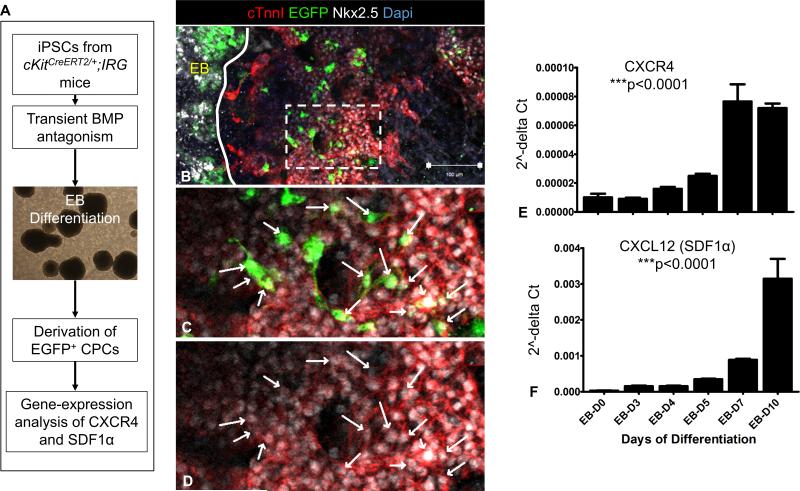
Figure 4. Expression of SDF1α and CXCR4 during derivation of CSCs from iPSCkit.
A, Schematic of the experimental design. B, Confocal immunofluorescence illustrates that the generation of CSCs from iPSCkit involves migration of CSCs from the embryoid bodies (EBs, solid line). C-D, high magnification of the area delineated by the inset in panel B, illustrates co-localization of EGFP with Nkx2.5 in the migrated CSC derivatives. E-F Gene-expression analysis during differentiation of iPSCkit to CSCs shows a dramatic upregulation in SDF1α and CXCR4. n=3; Scale bars, 100μm.

