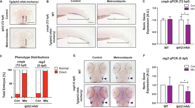
Figure 3. Selective Ablation of Serotonergic Neurons Decreases HSPCs.
(A) Serotonergic neurons were effectively ablated upon metronidazole (Mtz) treatment (10mM,12–72hpf) in tph2:nfsb-mcherry embryos as marked by tph2 WISH at 72hpf. Scale bar=200μM.
(B) Representative images of cmyb+ HSPCs in the CHT of WT and tph2:nfsb-mcherry fish after treatment as in (A). Scale bar=100μM.
(C) Expression of cmyb was significantly decreased by qPCR at 72hpf in tph2:nfsb-mcherry embryos with Mtz treatment, compared to control (n≥4 replicates).
(D) Qualitative phenotype distribution of cmyb and rag2 expression in tph2:nfsb-mcherry embryos at 72hpf and 5dpf, respectively.
(E) Representative images of rag2+ lymphoid cells in WT and tph2:nfsb-mCherry fish treated from 12hpf-5dpf with Mtz. Scale bar=200μM.
(F) Expression of rag2 was significantly decreased by qPCR in tph2:nfsb-mcherry embryos by Mtz treatment at 5dpf (n≥3 replicates).
mean±SD; two-way ANOVA, Holm-Sidak post hoc: *p<0.05.
See also Figure S4.

