Fig. 9.
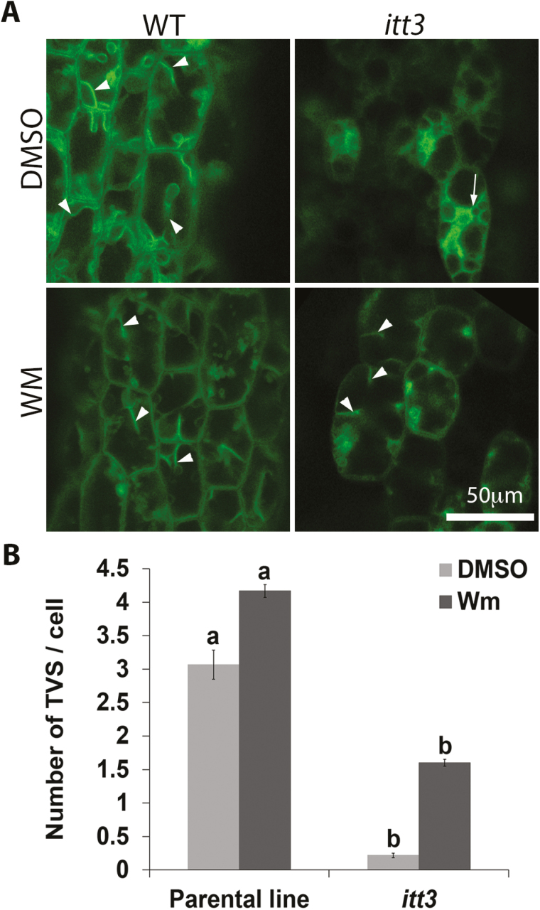
Wm treatment induces the formation of transvacuolar strands (TVSs). (A) Thick optical sections were captured for TVS quantification at low magnification. Example images used from WT and itt3 hypocotyls are shown for both DMSO and Wm treatments. The types of TVS that were counted are indicated with arrowheads. Note that membranes that fully surrounded a vacuole in itt3 were not included (arrows). (B) Average number of TVSs per cell in parental and itt3. n=60 cells total from four seedlings per genotype. Mean values are statistically significant (Student’s t test, two tailed, P≤0.05) if letters are different.

