Figure 1.
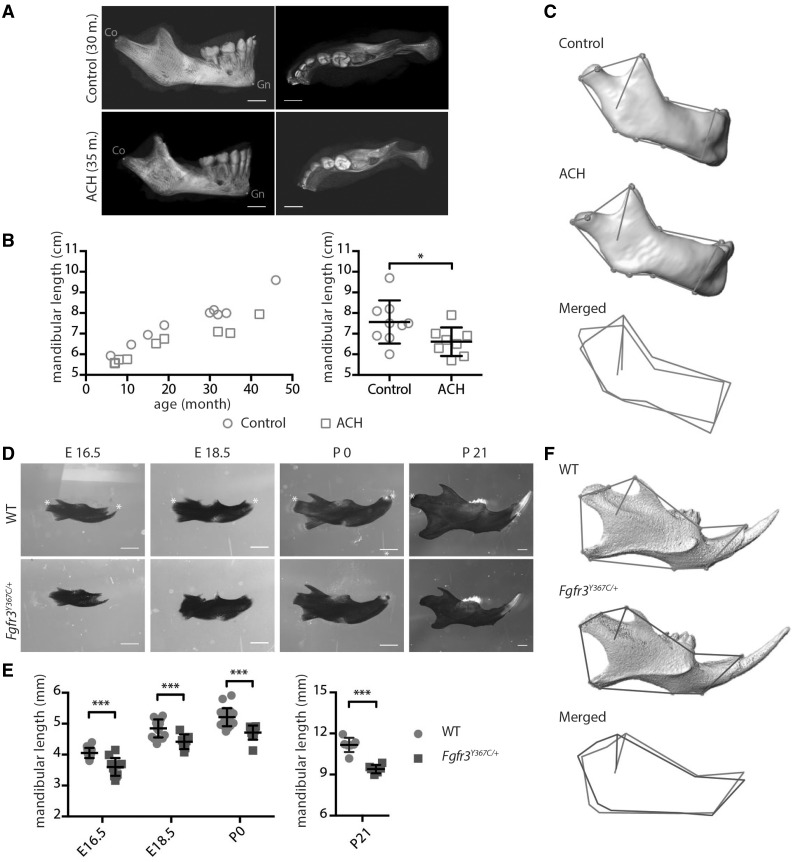
FGFR3 over activation in humans and mice results in mandibular hypoplasia and dysmorphogenesis. (A) 3D reconstructed CT images with volume rendering of a control and ACH patient (scale bar = 1 cm). (B) Mandible length, measured as the distance between condylion (Co) and gnathion (Gn) on sagittal sections of 3D reconstructed CT images of controls (n = 9, mean age: 24.9 months) or ACH patients (n = 8, mean age: 21.3 months) and plotted against the age of the children. (C) Landmarks and associated wireframes measured on the 3D reconstructed human mandibles and corresponding superimposition of the control and ACH wireframes computed on the basis of PC scores along PC1, the PC best separating the two groups and accounting for 47% of total shape variance. (D and E) Mandible length of embryos (E16.5 and E18.5), new born and 3-week-old WT and Fgfr3Y367C/+ mice (n ≥ 6 individuals for each age and genotype), measured following alcian blue and alizarin red staining between the condylar and symphyseal ends (marked with *) (scale bar = 1 mm). (F) Landmarks and associated wireframes measured on the 3D reconstructed mouse mandibles and corresponding superimposition of the control and Fgfr3Y367C/+ wireframes computed on the basis of PC scores along PC1, the PC best separating the two groups and accounting for 78% of total shape variance. Data shown as mean with SD; *P < 0.05, ***P < 0.005.

