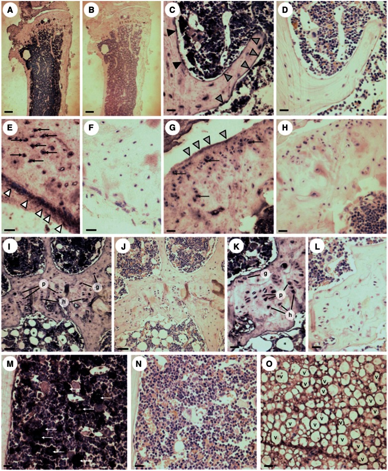
Figure 2.
Immunolocalisation of Opa3 in mouse tibiae. Immunohistochemical visualisation of Opa3 expression in 6-month-old WT tibiae with (A,C,E,G,I,K,M,O) and without (B,D,F,H,J,L,N) application of the primary rabbit anti-mouse Opa3 antibody, using the ImmPRESS peroxidase secondary antibody in conjunction with BCIP/NBT (5-bromo-4-chloro-3-indolyl phosphate/nitroblue terazolium) peroxidase substrate to produce indigo labelling, all sections being counterstained with haematoxylin and eosin. The immunoreactivity observed under low power (A) was located in osteoblasts on the endosteal (black arrowheads) and trabecular (grey arrowheads) (C), surfaces and on the surface of the cortical periostium (white arrowheads; E) and synovial cartilage (light grey arrowheads; G). In addition, Opa3 immunoreactivity was observed in cortical osteocytes (black arrows; E), synovial chondrocytes (dark grey arrows; G), germinal (g), proliferative (p) and hypertrophic (h) growth plate chondrocytes (I,K), mesenchymal stem cell rosettes (white arrows) in the mid-diaphyseal marrow (M) and adipocytes in the proximal (*; I,K) and distal (v;O) marrow (Scale bars: 300 µm (A,B), 50µm (C,D,I,J,M,N,O) and 20 µm (E,F,G,H,K,L).

