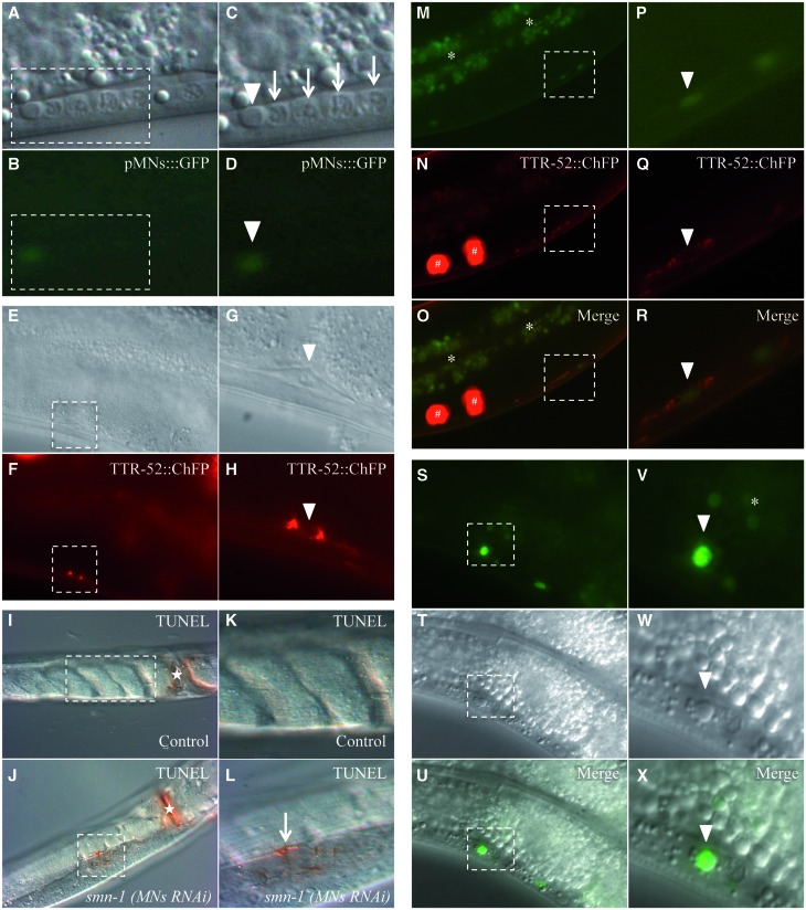
Figure 3.
The degeneration of motor neurons after smn-1 knock-down ends with the death of the cell and accumulation of fluorescence. The motor neuron death induced by smn-1(MNs RNAi) can be visualized in four different ways. (A–D) Using DIC microscopy in the ventral cord of smn-1 transgenic animals we observed a button-like structure typical of apoptotic dying cells (white arrowheads) in association with a neuron faintly expressing GFP from the oxIs12 (pMNs::GFP) transgene. Arrows correspond to viable neuronal nuclei in the ventral cord. (C, D) Enlargements of areas outlined in (A) and (B). Upper images were obtained with visible light, and lower images with GFP filter epifluorescence. (E–H) A TTR-52::ChFP positive neuron, with a typical ring shape, is observed in the ventral cord of smn-1(MNs RNAi), smIs119 (TTR-52::ChFP) double transgenic animals (white arrowheads). A quantification of TTR-52::ChFP positive neurons is reported in Supplementary Material, Table S7. (G, H) Enlargements of areas outlined in (E) and (F). Upper panels were obtained with visible light, and lower panels were obtained with Texas Red filter epifluorescence. (I–L) Apoptotic dying neurons in the ventral cord were analyzed in permeabilized animals using TUNEL assay. smn-1(MNs RNAi) knocked-down animals, but not controls, presented TUNEL-reactive cells along the ventral cord. (I) Control animal, adult stage, presenting no labeling in the cord. (K) Enlargement of the area outlined in I. (J) smn-1(MNs RNAi) animal, adult stage, presenting a positively labeled cell in the cord. (L) Enlargement of the area outlined in J, showing the labeled cell in the cord (arrow). n > 40 for control and for smn-1(MNs RNAi). White asterisks correspond to the vulva, which is always labeled. All images were taken with visible light and DIC. (M–R) Accumulation of fluorescence is sometimes surrounded by a TTR-52::ChFP ring (white arrowheads), in smn-1(MNs RNAi), smIs119 (TTR-52::ChFP) double transgenic animals. (P–R) Enlargements of areas outlined in (M)–(O). In the upper panels images were taken with epifluorescence using a GFP filter; in the middle panels a Texas Red filter was used, and the lower panels are merge of the two images. (S–X) Accumulation of fluorescence sometimes corresponds to a button-like structure, using DIC microscopy, typical of apoptotic dying cells (white arrowheads). A quantification of neurons accumulating fluorescence is reported in Figure 4A and in Supplementary Material, Table S7. (V–X) Enlargements of areas outlined in (S)–(U). In the upper panels images were taken with epifluorescence using a GFP filter; the middle panels are DIC images, and in the lower panels are merge of the two images. White asterisks correspond to intestinal autofluorescence and white hashtags to coelomocytes, scavenger cells that phagocyte secreted TTR-52::ChFP. Anterior is right and ventral is down in all images, except in (I)–(L) which are ventral views. All transgenic strains were obtained using an HC of interfering construct.

