Figure 4.
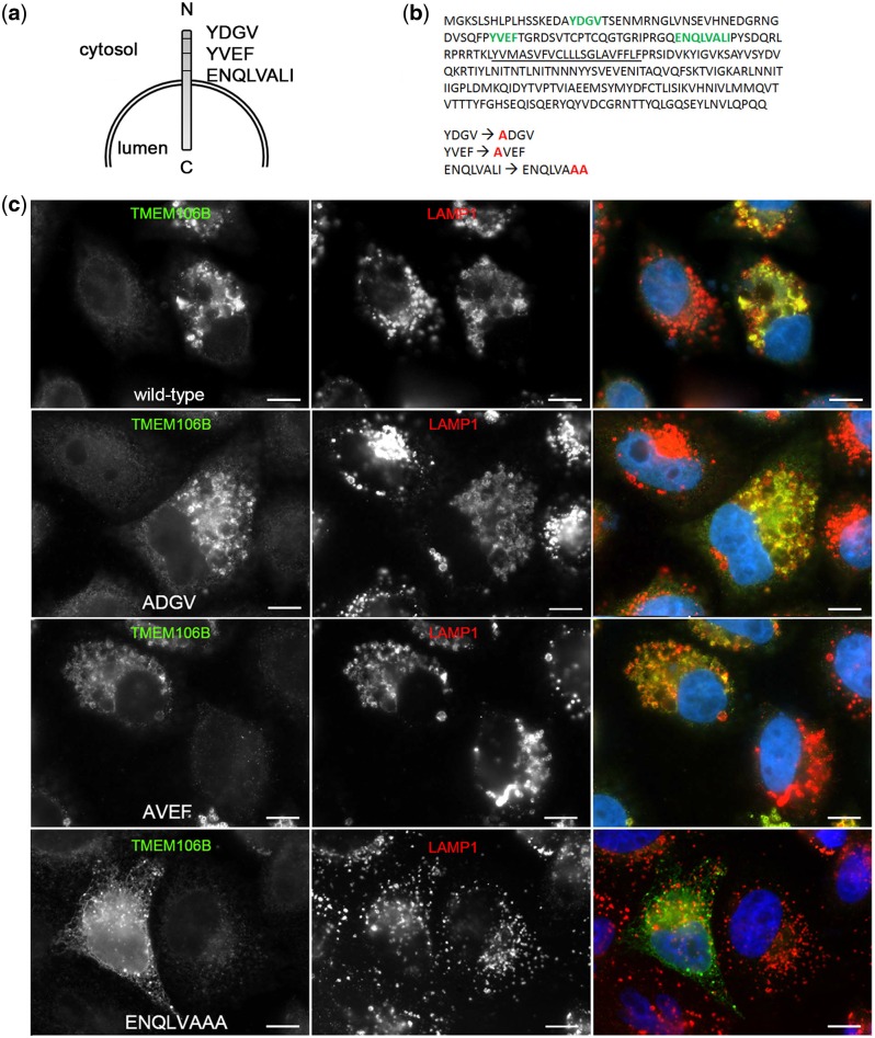
ENQLVALI is a potential lysosomal sorting motif for TMEM106B. (a) Three potential classical lysosomal targeting motifs—two tyrosine motifs and one isoleucine/dileucine motif—were identified in the N-terminal domain of TMEM106B. (b) The primary amino acid sequence of TMEM106B, with potential lysosomal targeting motifs depicted in green, is shown. Specific residues were individually mutated to alanine residues, with mutated residues indicated in red. (c) Double-label immunofluorescence microscopy demonstrates that wild-type TMEM106B (top row), ADGV-TMEM106B (second row) and AVEF-TMEM106B (third row) continue to co-localize strongly with LAMP1. In contrast, ENQLVAAA-TMEM106B (bottom row) appears diffusely throughout the cytoplasm of HeLa cells and exhibits decreased co-localization with LAMP1. For all constructs, the right-most panel shows merged channels for TMEM106B (green) and LAMP1 (red), with individual channels shown in monochrome in the left and middle panels. Scale bars = 10 µm. TMEM106B is detected by N2077 antibody.

