Figure 6.
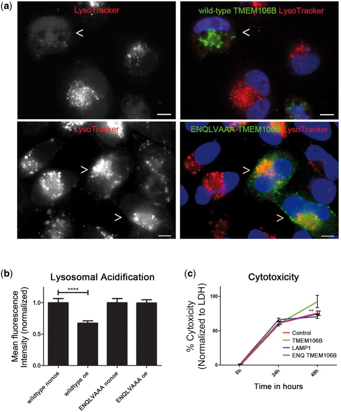
The effects of increased TMEM106B expression depend on proper localization of TMEM106B to lysosomes. (a, b) While expression of wild-type TMEM106B (FLAG-TMEM106B construct) in HeLa cells impairs lysosomal acidification (as demonstrated by decreased LysoTracker MFI, top row, arrowhead indicates TMEM106B over-expressing cell), expression of ENQLVAAA-TMEM106B does not significantly alter organelle acidification as compared with neighboring non-over-expressers (bottom row, arrowheads indicate ENQLVAAA-TMEM106B over-expressing cells). Representative images are shown in (a), and means ± SEM for six replicates performed on 3 days are shown in (b). Scale bars = 10 µm. Wild-type TMEM106B and ENQLVAAA-TMEM106B are detected by their FLAG tags in (a). (c) Cytotoxicity is rescued by mutation of critical residues within the potential dileucine lysosomal sorting motif (ENQLVALI to ENQLVAAA) in TMEM106B. While wild-type TMEM106B expression in HeLa cells induces cytotoxicity by 48 h (TMEM106B, green line), the loss of lysosomal localization (ENQ TMEM106B, pink) rescues this cytotoxicity to levels seen with transfection of vector only (control, red line, almost entirely overlapped by pink line). Cytotoxicity seen with expression LAMP1 (blue line) is also shown for comparison purposes; the LAMP1 data is repeated from Figure 3f. Beyond the 48-h time point, wild-type TMEM106B-expressing cells are largely lost from the culture medium due to cell death. % cytotoxicity is calculated in comparison to the maximal LDH release induced by treatment with Triton X as described in Materials and Methods section. Data are combined for six replicates performed on three different days. **p < 0.01. ****p < 0.0001.

