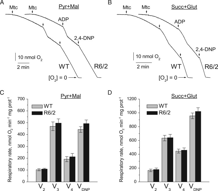
Figure 3.
Respiratory activity of brain synaptic mitochondria isolated from 6- to 8-week-old WT (thin traces) and R6/2 (thick traces) mice. In (A) and (B) are representative traces of mitochondrial O2 consumption for mitochondria at 37 °C in incubation medium supplemented with pyruvate (3 mM) plus malate (1 mM) or succinate (3 mM) plus glutamate (3 mM), respectively. Arrows indicate the addition of either synaptic mitochondria (Mtc), 200 µM ADP, or 60 µM 2,4-DNP. In (C) and (D) are statistical analyses of respiratory rates. Data are presented as mean ± SEM from five separate experiments.

