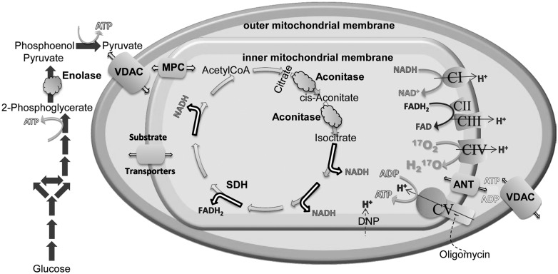
Figure 7.
Metabolic pathways and mitochondrial proteins altered or implicated in the altered function observed in this study. Changes in protein expression in enolase (round cloud) may slow glycolytic ATP production (glycolysis, solid grey arrows). Pyruvate, as well as other mitochondrial substrates, crosses the mitochondrial outer membrane when the voltage dependent anion channel (VDAC, large barrel) is open. Pyruvate is transported across the inner mitochondrial membrane through the mitochondrial pyruvate carrier (MPC). Other substrates utilize a variety of transporters (small barrels) to enter the mitochondrial matrix. Decreases in striatal VDAC expression may limit substrate availability. Inside the matrix, pyruvate enters the TCA cycle (open single headed arrows). Decreases in expression of cortical aconitase (oval cloud) may limit flux through TCA. These upstream alterations may limit flux through the electron transport chain complexes and oxidative phosphorylation, whose overall expression levels were largely unchanged. In vivo CMRO2 was measured from the conversion of 17O2 to O by complex IV (curved arrow). ATP produced by complex V and ADP cross the inner mitochondrial membrane through the adenine nucleotide transporter (ANT) and the outer mitochondrial membrane through VDAC. Decreases in striatal VDAC could also limit ATP and ADP movement into and out of mitochondria. The site of action of DNP and oligomycin are also indicated.

