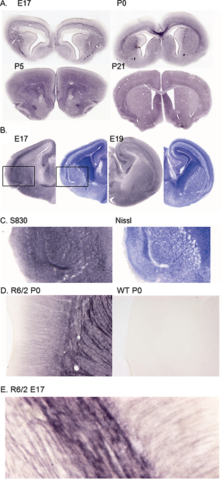
Fig.1.
A. S830 staining of R6/2 mouse brain at E17, P0, P5 and P21 showing staining initially restricted to developing white matter tracts and appearing extracellular. B. S830 (left) and Nissl (right) staining of YAC128 at E17 and E19 showing preponderance of S830 staining associated with developing white matter tracts and fiber bundles. C. Enlargement of areas shown in B: S830 (left) and Nissl (right) reveal inverse relationship between S830 and Nissl staining, particularly in the developing external capsule and fiber bundles in the developing striatum. D. Enlargement of a similar region in P0 R6/2 mouse brain showing diffuse staining of corticostriatal fibers with weak staining of cortical dendrites (left); staining of the same region in WT P0 brain (right) under identical conditions reveals the total absence of immunoreactivity with S830. E. High power view of external capsule of E17 R6/2 mouse brain stained with S830 showing diffuse nature of staining and complete absence of discrete mHTT aggregates; field size: 200×80 μ.

