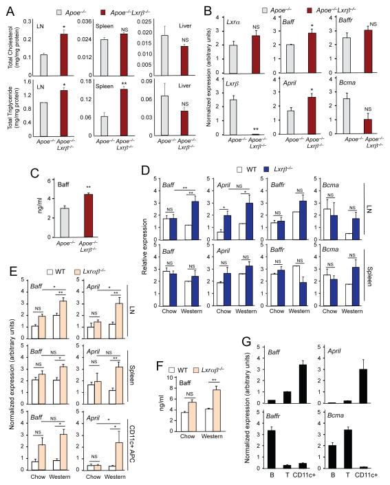
Figure 3. Cholesterol accumulation in lymphoid organs promotes the production of Baff and April.
(A) Lipid was extracted from lymph node, spleen and liver of 8-week-old Apoe−/− and Apoe−/−Lxrβ−/− mice. Total masses of cholesterol and triglyceride were determined by colorimetric methods. N=3. (B) Gene expression in lymph node of 8-week-old Apoe−/− and Apoe−/−Lxrβ−/− mice analyzed by real-time PCR. (C) Plasma Baff concentration in 8-week-old ApoE−/− and Apoe−/−Lxrβ−/− mice determined by ELISA. (D) Gene expression in lymph node (upper) and spleen (bottom) of wild-type and Lxrβ−/− mice fed chow or Western diet for 16 weeks analyzed by real-time PCR. (E) Gene expression in lymph node (upper), spleen (middle) and CD11c+ antigen-presenting cells (APC) (bottom) of wild-type and Lxrαβ−/− mice fed chow or Western diet for 12 weeks analyzed by real-time PCR. (F) Plasma Baff concentration in wild-type and Lxrαβ−/− mice fed chow or Western diet for 12 weeks determined by ELISA. (G) Pan B cells, pan T cells and APC were isolated from spleen of wild-type mice. Gene expression was analyzed by real-time PCR. N=4–6 per group. Statistical analysis was performed with Student’s t test (A–C) and two-way ANOVA (D–F). *p < 0.05, **p < 0.01, NS, not significant. Error bars represent means +/− SEM.

