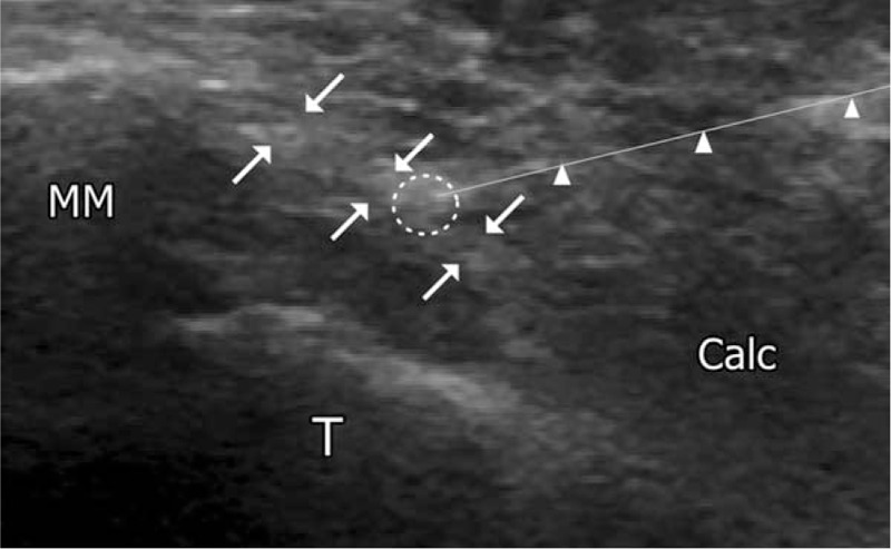
Figure 3.

Ultrasound image during injection of polydeoxyribonucleotide in the posterior tibial tendon. Arrowhead indicates the block needle. Calc = calcaneus, posterior tibial tendon (arrows), block needle (arrow head), injection site of polydeoxyribonucleotide (white circle), MM = medial malleolus, T = talus.
