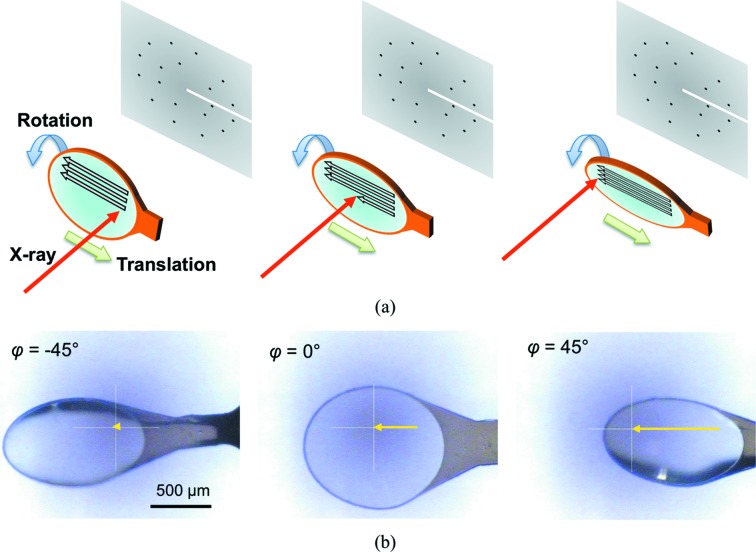
Figure 1.
Overview of SS-ROX data collection. (a) Schematic diagrams of two-dimensional raster scans with goniometer rotation. The loop on which the crystals were loaded was raster-scanned with rotation of the goniometer spindle axis. (b) Photographs of a horizontal helical scan; yellow arrows represent relative movement of the incident X-ray position. φ is the goniometer spindle angle.

