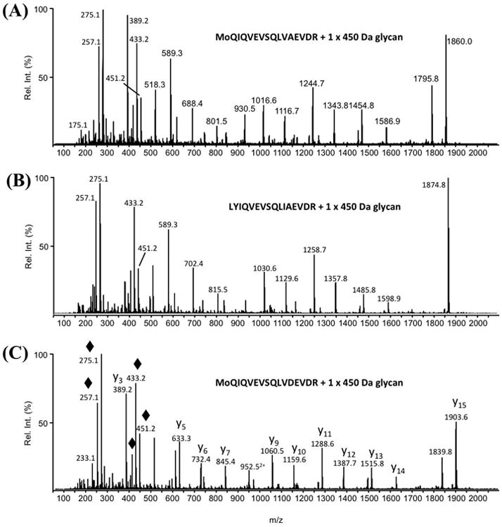
Fig. 3. Representative glycopeptide nLC-MS/MS spectra from three FlaB proteins, each modified with the 450.2 Da glycan.

(A) MS/MS spectrum of MH22+ ion at m/z 1155.6 from T108-123 of FlaB1, (B) MS/MS spectrum of MH22+ ion at m/z 1163.1 from T108-123 of FlaB2, and (C) MS/MS spectrum of MH22+ ion at m/z 1177.6 from T108-123 from FlaB3. The amino acid sequences of the three glycopeptides are presented in the insets. “Mo” indicates the presence of oxidized methionine. By way of example, the major peptide fragment ions are identified in the MS/MS spectrum presented in panel c (“y” fragment ions start at the C-terminal amino acids). The mass differences between the peptide fragment ions were used to determine the amino acid sequence of this glycopeptide. The lower region of each MS/MS spectrum is dominated by the m/z 451.2 glycan oxonium ion and its related fragment ions (indicated with ◆ in panel c) that arose from multiple losses of water as well as the loss of the hexanoyl moiety.
