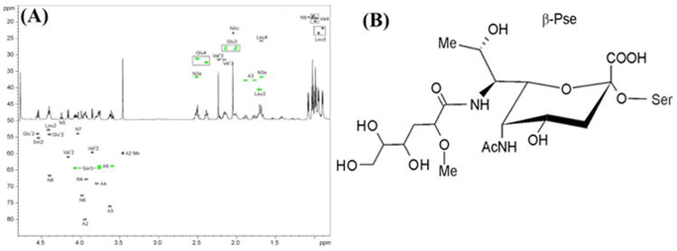
Fig. 5. 1H-13C HSQC spectrum of the glycopeptide from T. denticola and its 1H NMR spectrum.

(A) CH signals are black, and CH2 are green. Nonulosonic acid signals are labeled as N. (B) Structure of the flagellar glycan with Pse configuration (L-glycero-L-manno) of the nonulosonic acid residue β-linked to serine (NB C-8 may have the 8-epi-Pse configuration).
