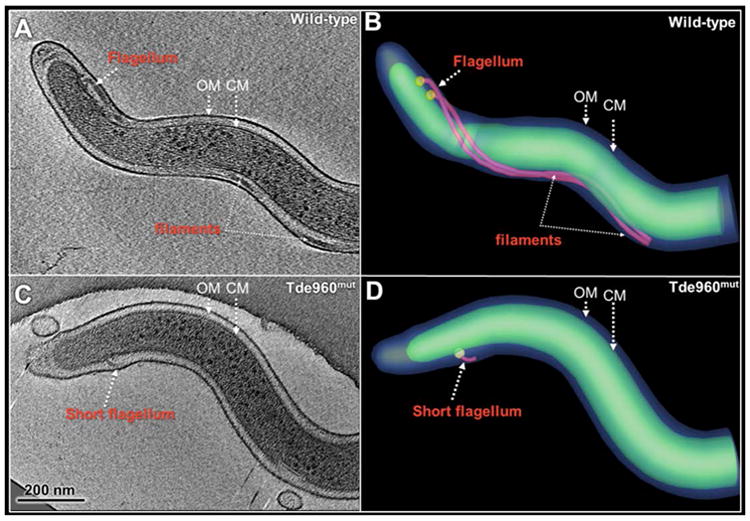
Fig. 8. Cryo-ET analysis of the wild-type and Tde960mut strains.

(A) A central slice of a typical tomographic reconstruction from a wild-type cell tip. PFs are observed in the periplasmic space between the outer membrane (OM) and the cytoplasmic membrane (CM). (B) The 3-D surface rendering of the reconstruction (A) shows two long PFs (colored in red) surrounding the cell body (green). (C) A central slice of a tomographic reconstruction from a Tde960mut cell. A short flagellum is attached to the CM. (D) The short flagellum is clearly shown in the surface rendering.
