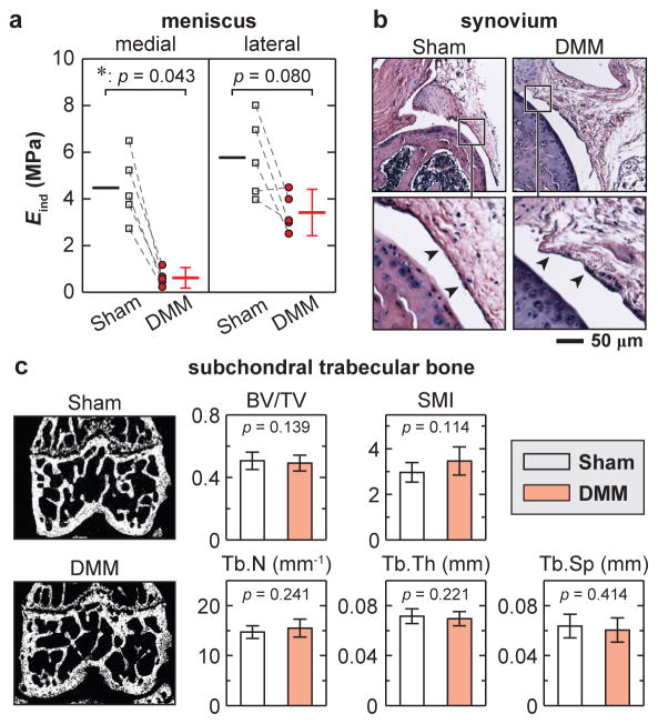
Fig. 6.
Osteoarthritic changes in meniscus, synovium and subchondral trabecular bone at 8 weeks after DMM surgery. a) Meniscus: AFM-based nanoindentation on meniscus surfaces illustrated marked modulus reduction on the medial side, and marginal changes on the lateral side after DMM (n = 5, each error bar represents 95% CI). b) Synovium: hematoxylin and eosin (H&E) staining showed no appreciable morphological differences (n = 4). c) Subchondral trabecular bone, left panel: frontal plane of μCT images, right panel: trabecular structural parameters calculated from μCT images (n = 10). Neither panel exhibited significant differences between DMM versus Sham knees.

