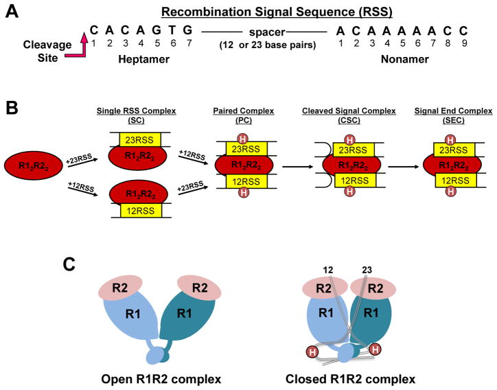
Figure 1. RAG-RSS complexes formed during V(D)J recombination.
(A) Consensus heptamer and nonamer sequences in the RSS. Only the DNA strand initially nicked by the RAG proteins is shown. Nicking occurs 5′ to the heptamer at the position indicated. (B) RAG complexes formed in the V(D)J recombination reaction. R1 and R2 refer to RAG1 and RAG2, respectively. Circles labeled H (in the PC, CSC, and SEC) refer to HMGB1 or HMGB2. (C) Cartoons representing structural models of R12R22 complexes. The DNA-free ‘apo’ and 12/23-RSS-bound R12R22 complexes are proposed to be in “open” and “closed” conformations, respectively (18).

