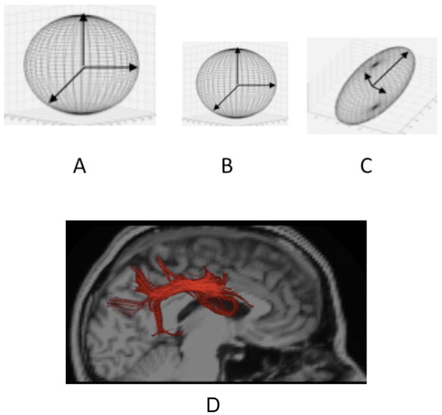
Figure 10.
Diffusion Tensors: A: Illustration of tensor from region with high isotropic diffusivity, as in cerebrospinal fluid. B: Tensor exhibiting isotropic, but lower diffusivity, as in gray matter. C: Elongated tensor exhibiting anisotropy, as in fiber tracts. D. Illustration of tractography of the superior longitudinal fasciculus (a major fiber tract connecting posterior with frontal parts of the cortex) shown in red.

