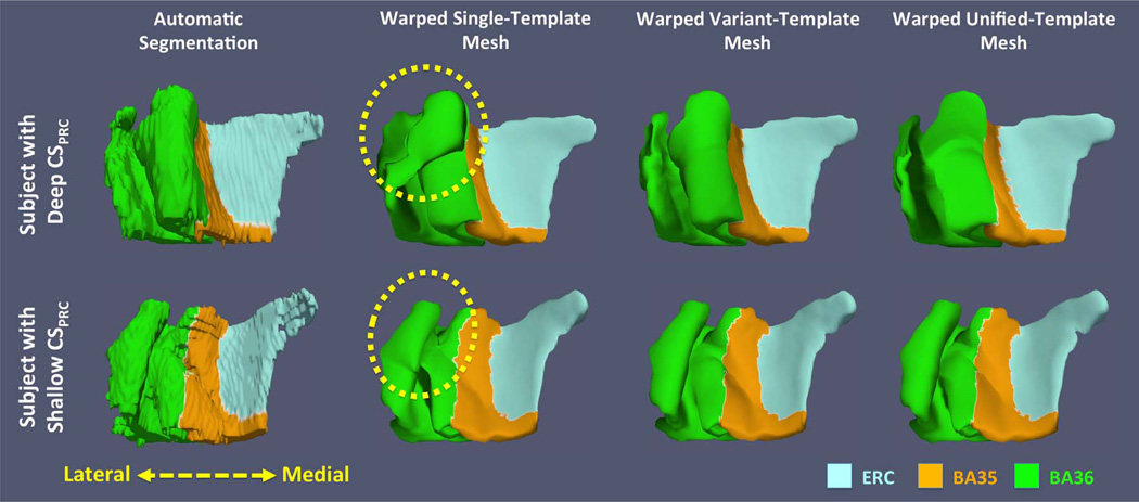
Fig. 8.
Comparisons of fitted smooth meshes from single-template, variant-template and unified-template approaches for subjects with deep (top) and shallow (bottom) CSPRC. The yellow dashed circles highlight the incorrect correspondences between subjects established by the single-template approach: the biological implausible structure in left single template (white arrow in Fig. 7) maps to the medial border of BA36 of one subject (top) and the lateral border of BA36 of the other subject (bottom). (For interpretation of the references to color in this figure legend, the reader is referred to the web version of this article.)

