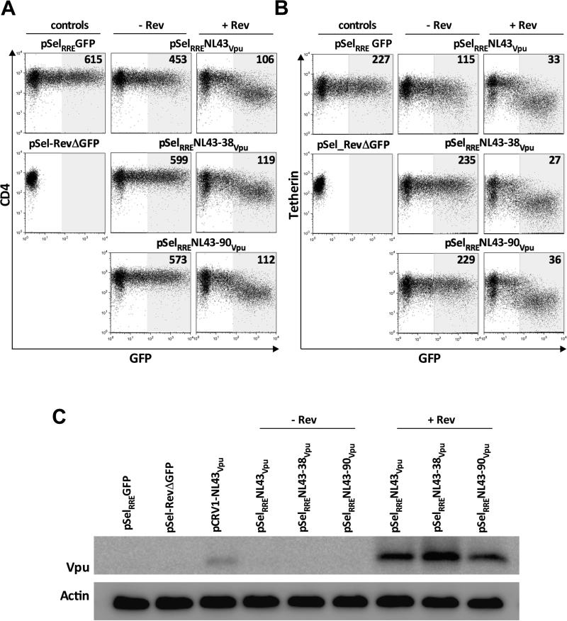
Figure 3. Robust Vpu function/expression, regardless of the presence of upstream bases, using a Rev/RRE-dependent system.
Panel A: Representative flow cytometry plots of Vpu-mediated CD4 downregulation by control (left column) and NL43 Vpu sequences cloned into pSelRRE-GFP, cotransfected without (middle column) and with (right column) a plasmid encoding HIV-1 Rev. Controls pSELRRE-GFP and pSel_RevΔGFP are shown separately to demonstrate GFP expression from the former but not the latter. Numbers on flow plots represent the MFI of CD4 staining in shaded gate. Experiments were performed by transfecting 5 μg Vpu with 7 μg Rev DNA into 2.5 million cells. Panel B: same as A, except for tetherin downregulation. Panel C: Detection of Vpu levels from pSELRRE-GFP constructs, with or without Rev, by western blot, with actin as the housekeeping control.

