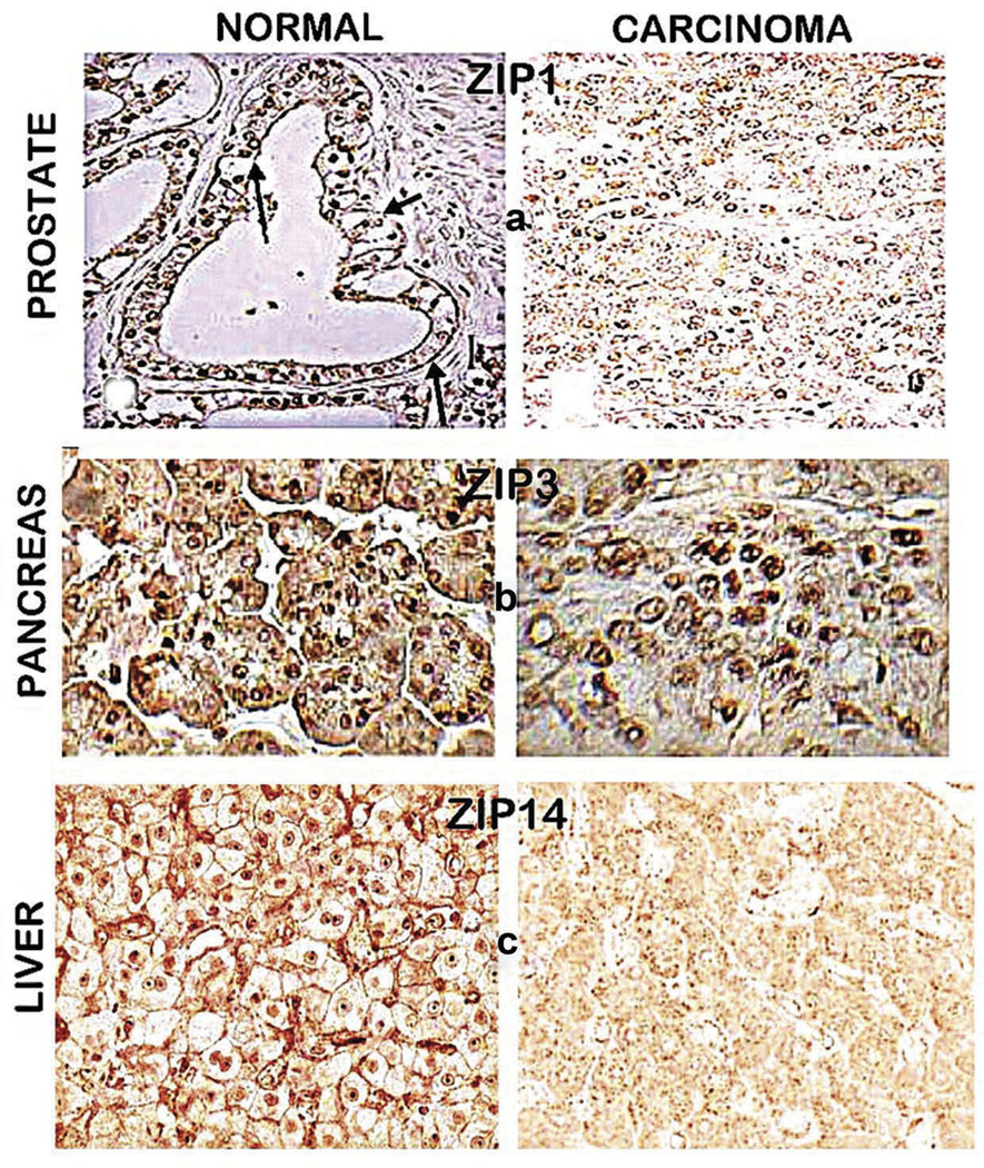
Figure 4.
ZIP transporter immunohistochemistry of normal versus cancer tissue sections. (a) Normal prostate acinar epithelium shows ZIP1 transporter localized at the basolateral and apical membrane; and absence of detectable membrane-localized transporter in malignant cells. Reproduced from [21]. (b) Normal pancreas shows ductal and acinar epithelium with ZIP3 localized at the basilar membrane; and the malignant cells with the absence of detectable plasma membrane localized ZIP3. Reproduced from [17]. (c) Normal liver shows hepatocytes with plasma membrane localized ZIP14; and hepatoma cells with absence of detectable plasma membrane ZIP14. Reproduced from [18].

