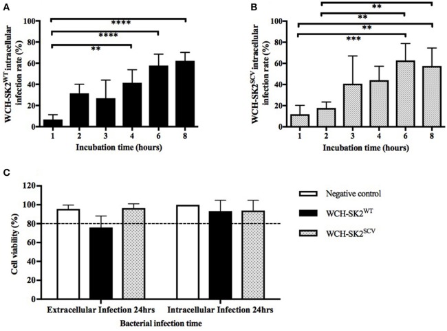
Figure 3.
WCH-SK2WT and WCH-SK2SCV intracellular infection rate and cell viability post infection. Comparison of intracellular infection rate in NuLi-1 cells between S. aureus WCH-SK2WT (A) and WCH-SK2SCV (B) strains (MOI 100). NuLi-1 cells were incubated with either WCH-SK2WT and WCH-SK2SCV for a variable incubation time. The cells were treated with lysostaphin to remove any extracellular S. aureus and the incubation was continued with gentamycin. The intracellular infection rate was determined 24 h post lysostaphin treatment. The data shown is mean ± SD of three independent experiments measured. **p ≤ 0.01, ***p ≤ 0.001, ****p ≤ 0.0001, Kruskal–Wallis test. NuLi-1 cell viability post extracellular and intracellular S. aureus infection was determined using an LDH assay (C). For extracellular infections, the S. aureus was incubated with the cells for 24 h. For intracellular infections, the S. aureus was incubated with the cells for 6 h followed by lysostaphin treatment. The cells were incubated with gentamycin for another 17.5 h for a total of 24 h of incubation respectively. The data shown is mean ± SD of three independent experiments measured. Kruskal–Wallis test. LDH = Lactate dehydrogenase, WCH-SK2WT = Staphylococcus aureus strain WCH-SK2 wild type. WCH-SK2SCV = Staphylococcus aureus strain WCH-SK2 small colony variant.

