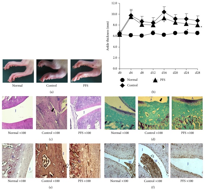
Figure 1.
PFS prophylactically suppress inflammation in CFA-induced arthritis in rats. AIA was induced as detailed in Methods. Rats were randomly divided into 3 treatment groups: normal, control, and PFS. PFS were administered orally until the day before tissue harvesting 28th day. (a) Representative photographs of days 21–27 paws from the 3 treatment groups. (b) Change in ankle thickness. The changes in paw thickness were measured every four days. The results are expressed as the mean ± standard error (n = 7–9). Statistical values conducted on days 0–28 for changes in paw thickness compared with control were as follows: ∗ p < 0.05 compared with the control group, Dunnett's test. ((c)–(f)) Representative photographs of knee joint tissues stained with safranin-O-fast green, toluidine blue-fast green, TRAP, or MMP-11 (magnification, 100x). Note that the intense synovial inflammatory infiltration ((c) black arrowhead), pannus formation ((c) arrowheads), cartilage destruction, extensive proteoglycan depletion ((d) black arrowhead), and MMP-11 infiltration ((f) arrowhead) were significantly reduced in the joints of PFS-treated rats compared with the respective control rats. A: articular cartilage; G: growth plate; J: joint space; M: synovial membrane; P: pannus formation.

