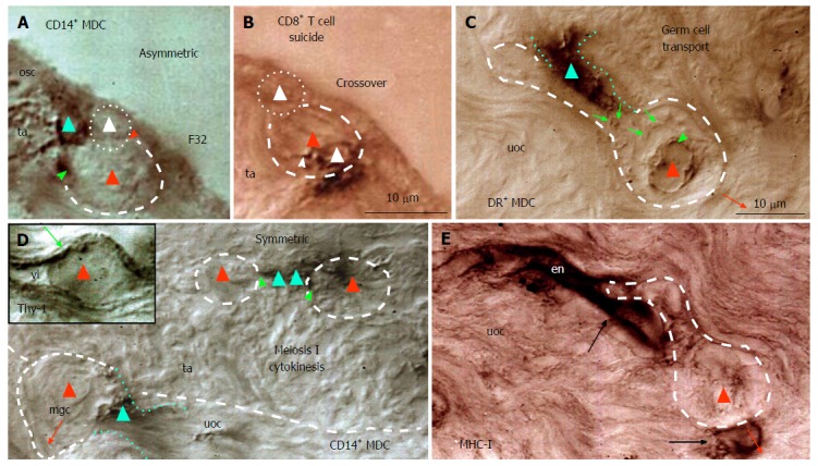
Figure 9.

Origin and migration of germ cells during the midfollicular phase in the adult human ovary. A: In the presence of primitive MDC (green triangle) the dividing ovarian stem cell (white triangle) produces a germ cell (red triangle); B: This is accompanied by the presence of CD8 T cell (white triangle) with extensions (arrowhead) within the germ cell; C: Migrating germ cell (dashed line) is accompanied by DR+ MDC (dotted lines), which releases DR (arrows) accumulating at the germ cell nucleus (arrowhead); D: In the tunica albuginea (ta) MDCs (green triangles) are associated (green arrowheads) with meiosis I cytokinesis (red triangles) and accompany moving germ cell (mgc) in the upper ovarian cortex (uoc). Inset shows Thy-1+ cortical venule containing germ cell (red triangle); E: Migrating germ cell lacking MHC-I expression is associated with stained endothelial cells of a venule in the upper ovarian cortex. F32 indicates a female patient’s age[18]. MDC: Monocyte-derived cell.
