Figure 2.

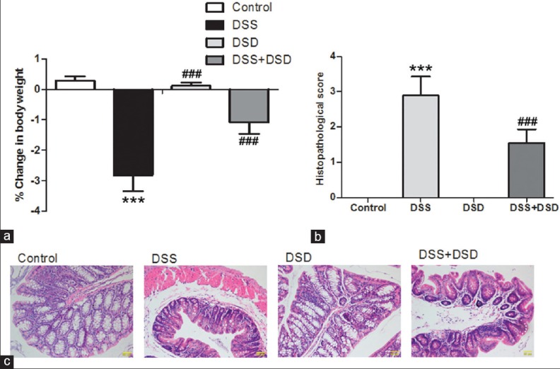
Deoxyschizandrin attenuated DSS-induced UC in mice. (a) Changes in the body weight of various groups of mice before and after the experiment. Histopathological scores (b) and HE staining (c) were used to evaluate the morphological changes in mouse colon tissues after DSS induction with and without deoxyschizandrin. The figure shows the representative results from repeated experiments (n = 6). Data are expressed as mean ± standard deviation. Compared with the Control group, ***P < 0.001; compared with the DSS group, ###P < 0.001
