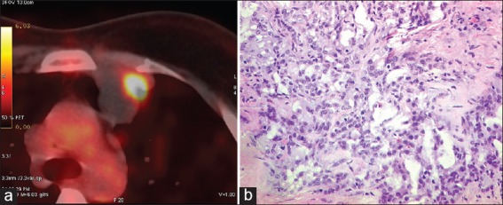
Figure 1.

(a) with positron emission tomography - computed tomography scan fluorodeoxyglucose (FDG) avid an anterior mediastinal node in pre-vascular region measuring (2.8 cm × 2.7 cm) with central necrosis and a focal area of increased FDG uptake. (b) Microphotograph showing infiltrating duct carcinoma, grade III, amidst sclerotic stroma (H and E, original magnification, ×200)
