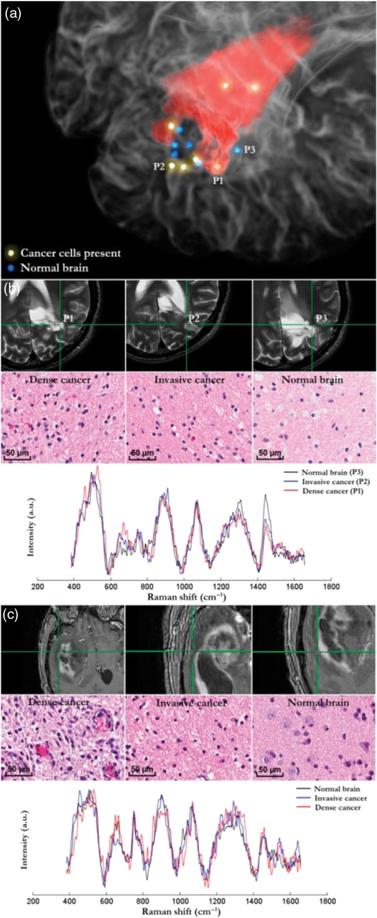
Fig. 2.
Raman spectroscopy measurements colocated on preoperative MRI-grade 2 and 4 gliomas. (a) 3-D rendering of a preoperative T2-weighted MRI overlaid with the segmentation of a grade 2 astrocytoma in red. Samples for each tissue type are indicated by P1, P2, and P3. (b) Pathology images for regions P1 to P3 in (a). P1, P2, and P3 are dense cancer, invasive cancer, and normal brain, respectively. The acquired Raman spectra are shown below. (c) In a different patient with grade 4 GBM, locations for dense cancer, invasive cancer, and normal brain are shown on a T1-weighted MRI with corresponding histopathology.

