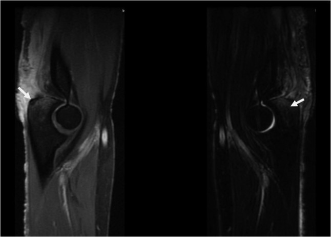
Figure 2:

Sagittal T1 (left) and T2 (right) magnetic resonance imaging of the right elbow showing inflammation around the triceps insertion with lytic lesion of the olecranon process of the ulna indicating osteomyelitis. Left arrow points to the lytic lesion in the olecranon process. Right arrow points to T2 hyperintense signal representing bone marrow edema.
