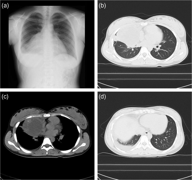
Figure 1:
Chest X-ray and CT at the initial visit. The chest X-ray showed a large infiltration in the right lower thorax (a). The CT scan showed a large mass with low-level pleural effusion, an internal fluid component and external parenchyma mixed with calcification in the anterior mediastinal area (b–d).

