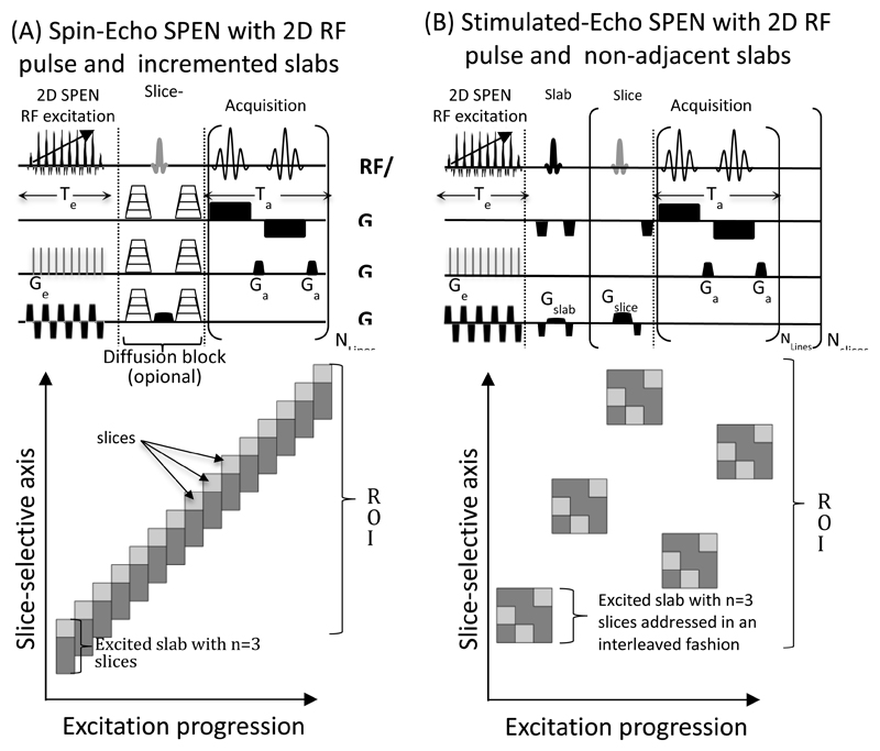
Figure 1.
Pulse sequences utilized in this study, incorporating the encoding of single slabs (in dark grey) by 2D spatial/spatial SPEN pulses, and subsequent single-shot acquisitions on single slices (lighter grey, illustrated for a case where n=3 slices fit per slab). (A) Spin-echo version, incorporating (optional) stepped gradients for diffusion-encoding. (B) Stimulated-echo version. This slab/slice operation was adopted in (A) owing to the scanner’s inability to excite sufficiently narrow slices; the targeted slice is thus selected by the 180˚ spin-echoing pulse. Notice that the incremented positions arrangements allows one to operate without saturation and/or interslab excitation delays; for these cases, the number of slabs excited equals the number of slices observed. The interleaved spatial disposition strategy adopted in (B) is meant to minimize “leakage” effects between consecutive excitations; in this case n slices are observed per excited slab.

